
Sources & acknowledgments
Credits.
Research figures
WiCAT, Figure 1: Mohammad Hosseini, Eray Erturk, Saba Hashemi, Maryam M. Shanechi, ICML (2026). Figure and abstract licensed under CC BY 4.0. The complete figure is resized and converted to WebP. Abstract whitespace and punctuation are normalized. Figure source. Abstract source.

BaRISTA, Figure 1: Lucine L. Oganesian, Saba Hashemi, Maryam M. Shanechi, NeurIPS (2025). Figure and abstract licensed under CC BY 4.0. The complete figure is resized and converted to WebP. Abstract whitespace and punctuation are normalized. Figure source. Abstract source.

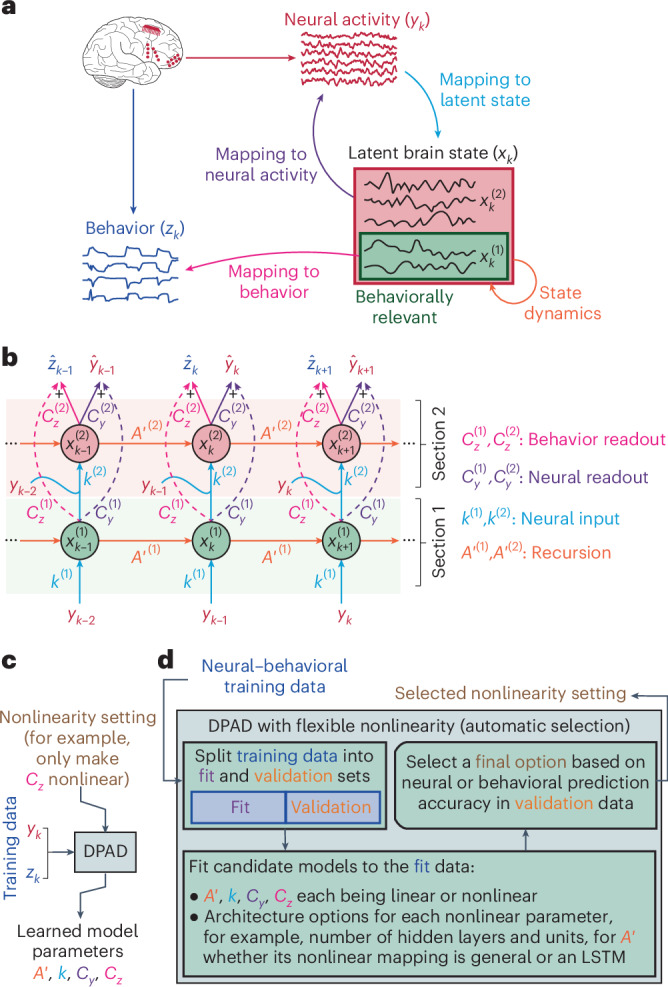
DPAD, Figure 1: Omid G. Sani, Bijan Pesaran, Maryam M. Shanechi, Nature Neuroscience (2024). Figure and abstract licensed under CC BY 4.0. The complete figure is resized and converted to WebP. Abstract whitespace and punctuation are normalized. The homepage preview is cropped to panel a; Research retains the full figure. Figure source. Abstract source.
DFINE, Figure 1: Hamidreza Abbaspourazad, Eray Erturk, Bijan Pesaran, Maryam M. Shanechi, Nature Biomedical Engineering (2024). Figure and abstract licensed under CC BY 4.0. The complete figure is resized and converted to WebP. Abstract whitespace and punctuation are normalized. Figure source. Abstract source.

SBIND, Figure 1: Sayed Mohammad Hosseini, Maryam Shanechi, ICML (2025). Figure and abstract licensed under CC BY 4.0. The complete figure is resized and converted to WebP. Abstract whitespace and punctuation are normalized. Figure source. Abstract source.

MRINE, Figure 1: Eray Erturk, Maryam M. Shanechi, NeurIPS (2025). Figure and abstract licensed under CC BY 4.0. The complete figure is resized and converted to WebP. Abstract whitespace and punctuation are normalized. Figure source. Abstract source.

BRAID, Figure 1: Parsa Vahidi, Omid G. Sani, Maryam M. Shanechi, ICLR (2025). Figure licensed under CC BY 4.0. The complete figure is resized and converted to WebP. Figure source.

IPSID, Figure 1: Parsa Vahidi, Omid G. Sani, Maryam M. Shanechi, PNAS (2024). Figure licensed under CC BY-NC-ND 4.0. The complete figure is resized and converted to WebP. Figure source.

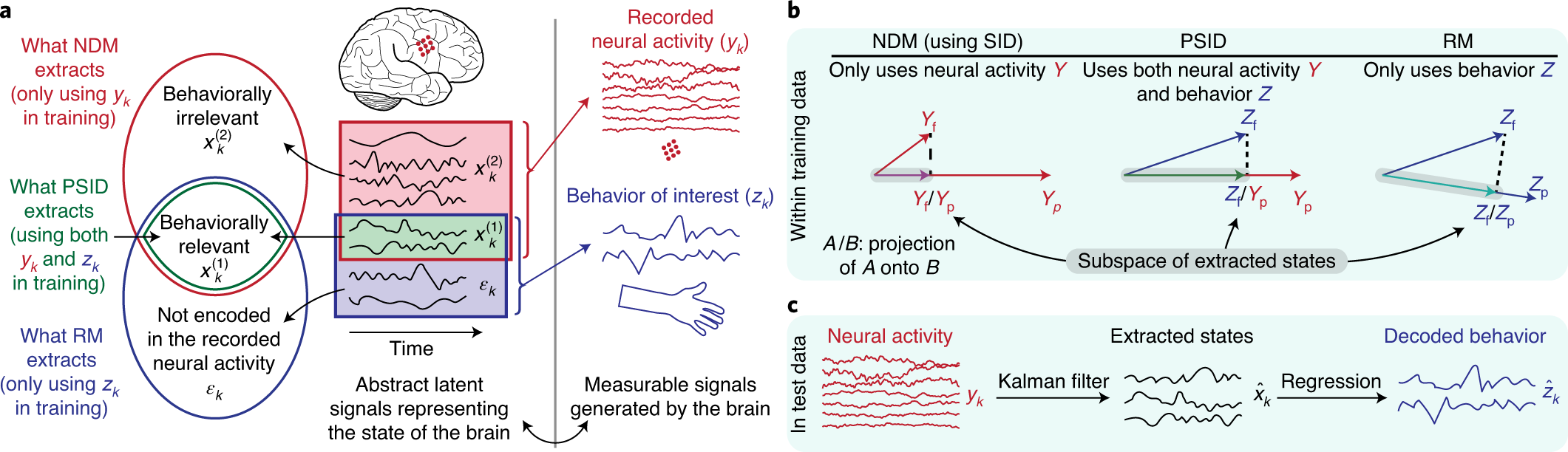
PSID, Figure 1: Omid G. Sani, Hamidreza Abbaspourazad, Yan T. Wong, Bijan Pesaran, Maryam M. Shanechi, Nature Neuroscience (2020). © 2020 The authors, under exclusive licence to Springer Nature America, Inc. The complete figure is resized and converted to WebP. Figure source.
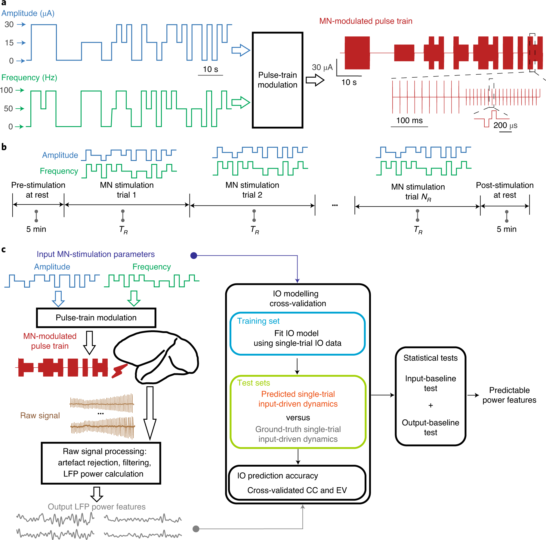
Dynamic brain stimulation, Figure 1: Yuxiao Yang, Shaoyu Qiao, Omid G. Sani, J. Isaac Sedillo, Breonna Ferrentino, Bijan Pesaran, Maryam M. Shanechi, Nature Biomedical Engineering (2021). © 2021 The authors, under exclusive licence to Springer Nature Limited. The complete figure is resized and converted to WebP. Figure source.
Journal covers
Nature Neuroscience, October 2024. Image: Ella Maru Studio, Omid Sani, Maryam Shanechi. Cover design: Marina Spence Cover artwork is reproduced from the lab’s existing cover collection, resized and converted to WebP without cropping. Journal-cover credits are separate from the article licenses above.
Nature Biomedical Engineering, January 2024. Image: Ella Marushchenko and Ekaterina Zvorykina (Ella Maru Studio, Inc.). Cover design: Alex Wing. Cover artwork is reproduced from the lab’s existing cover collection, resized and converted to WebP without cropping. Journal-cover credits are separate from the article licenses above.
Nature Biomedical Engineering, April 2021. Image: Ella Marushchenko and Kate Zvorykina (Ella Maru Studio); concept: Maryam Shanechi, Yuxiao Yang, Omid Sani (University of Southern California). Cover Design: Alex Wing. Cover artwork is reproduced from the lab’s existing cover collection, resized and converted to WebP without cropping. Journal-cover credits are separate from the article licenses above.
Nature Biotechnology, October 2018. Image credit: Ella Maru Studio; Omid Sani, Yuxiao Yang and Maryam Shanechi Cover artwork is reproduced from the lab’s existing cover collection, resized and converted to WebP without cropping. Journal-cover credits are separate from the article licenses above.
Bibliography & talks
The publication list preserves the lab’s bibliography, with research topic tags added for this site. Superseded preprint entries are merged with the final papers; distinct conference and journal publications remain separate. Citations without a verified publication link remain available as text.
Talk titles, dates, event details, and recording links were migrated from the lab’s previous website. Thumbnails use the original YouTube video images or the lab’s existing recording cards, converted to WebP without cropping.
The news archive preserves the lab’s dated announcements, with concise titles and summaries for this website.
Identity & illustrations
Visualization tap feedback uses ios-haptics by tijn, under the MIT license.
The navigation lens uses liquidGL by NaughtyDuk, adapted from its menu-bar demo and distributed under the MIT license.
The lens animation uses GSAP and the menu-bar demo’s elastic easing, under the GSAP Standard License.
People biographies, profile links, portraits, and Maryam Shanechi’s titles were migrated from the lab’s previous website. Portraits are locally hosted as WebP images. Join us adapts the lab’s existing open positions content.
USC logotype from USC Identity. Source Sans 3 is locally hosted under the SIL Open Font License.
The motion graphics, interface icons, and research illustrations were created for this website. The interactive values are synthetic rather than trained-model outputs.
Brain anatomy
The imaging illustration uses cortical boundaries derived from the Allen Mouse Brain Common Coordinate Framework, CCFv3 (2017), 100 µm annotation, Allen Institute for Brain Science. We projected the cortical annotation dorsally, combined layer labels, simplified contours, and recolored the regions. The animated activity and subject variations are illustrative. See Wang et al., Cell (2020), the Allen Institute noncommercial terms, and its citation policy.
The brain surface and markers use both hemispheres of the FreeSurfer fsaverage cortical template, distributed with Nilearn's fsaverage5 surfaces. The original meshes were packaged and quantized for this scene; markers sample triangle centers across both hemispheres. It is an average anatomical template, not an individual participant's MRI.
Source notices and terms: FreeSurfer license, Nilearn BSD license. Source provenance and modifications.
The shaded surface is rendered with Three.js, released under the MIT License.